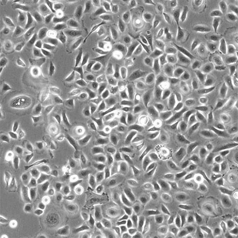
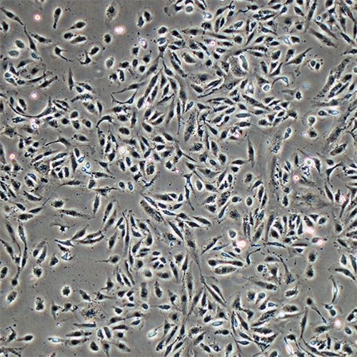
Human eye cells on a chip.
We screen eye drops, drugs and devices on living human eye cells under real dry eye stress, with quality control that makes results reproducible in any lab.
Better eye products start with better data.
Real human eye cells
Corneal and conjunctival cells grown on a cell culture plate or on a microfluidic chip that recreates flow, the tear film and dry eye disease.
Quality control
Our imaging algorithm checks the quality of every cell culture test. It is designed for human corneal and conjunctival cells, so results are standardized across studies.
Screening and benchmarking
We test and compare eye drops, drugs and devices for industry and academic partners in preclinical studies, before animal studies.
Watch cells respond in real time
Cells change all the time. Our chips work with standard lab microscopes for live-cell time-lapse imaging, and our image analysis turns every frame into data on how cells respond.